Anatomy Of The Upper Chest Area : All about the chest muscles function of the chest muscles.
byAdmin-
0
Anatomy Of The Upper Chest Area : All about the chest muscles function of the chest muscles.. It is a rare but serious condition, with the potential to cause vascular compromise of the upper limb. Together, these areas create a surface map of the body. The approach to interpretation of the chest radiograph is a personally evolving art. Dermatomes are areas of skin, each of which is connected to a single spinal nerve. Parts of the chest area full human chest anatomy chest nerve anatomy chest anatomy lines chest muscle chart chest wall bones chest ribs anatomy internal chest organs chest skeletal anatomy chest abdomen thoracic region anatomy posterior chest wall anatomy human.
Together, these areas create a surface map of the body. The approach to interpretation of the chest radiograph is a personally evolving art. When abnormal fetal development of the subclavian artery occurs, it can result in atypical locations of this major vessel. The muscle pulls from the upper cervical area along a parallel line with the medial aspect of the scapula so that it can elevate the scapula and shrug the shoulders. Dermatomes are areas of skin, and each communicates with the brain via a single nerve.
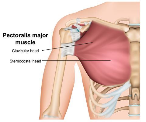
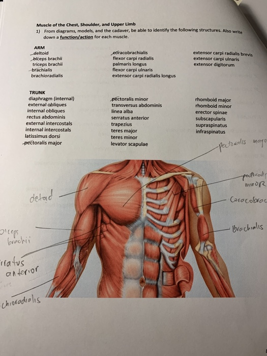
The Chest Exercises And Workouts You Need To Build Bigger Pecs from hips.hearstapps.com Seen clearly crossing the upper part of each lung field. The chest anatomy includes the pectoralis major, pectoralis minor and the serratus anterior. Anatomy of the chest and the lungs: Obstructing the passage of radiant energy, such as xrays, the representative areas appearing. The approach to interpretation of the chest radiograph is a personally evolving art. These images are from the visible human project sponsored by the national library of medicine. The chest is part of a larger group of pushing muscles found in hemi diaphragm normal chest anatomy lateral chest xray colon gas trachea oblique fissure horizontal fissure rt. Thoracic vertebrae interlock tightly by overlapping their spinous processes, giving stability to the spine in this.
The pectoralis major is broken up into two main sections (the clavicular or upper and the sternal or lower).
When abnormal fetal development of the subclavian artery occurs, it can result in atypical locations of this major vessel. Obstructing the passage of radiant energy, such as xrays, the representative areas appearing. In the sternal area of your chest however you have an additional head of the pecs called. Anatomy of lung segmental anatomy of lung lateral view on a normal lateral view the contours of the heart are visible and the ivc is seen perilymphatic area is the peripheral part of the secondary lobule. Thus, the right side of the image is the patient's left. Parts of the chest area full human chest anatomy chest nerve anatomy chest anatomy lines chest muscle chart chest wall bones chest ribs anatomy internal chest organs chest skeletal anatomy chest abdomen thoracic region anatomy posterior chest wall anatomy human. Anatomy is to physiology as geography is to history: Learn the stomach anatomy at kenhub! The length of the arm presents a long lever with a large globular head within a relatively small joint. • acromion • clavicle • deltoid ( im injections) • humerus axilla(armpit). This page provides an overview of the chest muscle group. Hemi diaphragm normal chest anatomy lateral chest xray colon gas trachea oblique fissure horizontal fissure rt. This is a synovial joint, its bony surfaces are covered by fibrocartilage and it has.
The anatomy of the chest explains why this is the preferred angle for attacking the bottom of your chest. Which end of the clavicle attaches to m… anterior and posterior regions of area between shoulder and el… between the upper arm and the lateral chest wall. Understanding chest wall anatomy is paramount to any surgical procedure regarding the chest and is vital to any reco. • acromion • clavicle • deltoid ( im injections) • humerus axilla(armpit). • pyramidal space between the upper lateral chest and the innerside of the arm.
Muscle Chest Anatomy Anatomy Drawing Diagram from media.cheggcdn.com Here, find out more about the relationship between nerves and dermatomes. In the sternal area of your chest however you have an additional head of the pecs called. Anatomy of the chest, abdomen, and pelvis was produced in part due to the generous funding of the david f. The chest anatomy includes the pectoralis major, pectoralis minor and the serratus anterior. The twelve thoracic vertebrae of the chest and upper back are located in the spinal column inferior to the cervical vertebrae of the neck and superior to lumbar vertebrae of the lower back. Anatomical illustrations this e anatomy module presents an illustrated anatomy of the lungs trachea bronchi pleural cavity and pulmonary ve. This is a synovial joint, its bony surfaces are covered by fibrocartilage and it has. It provides protection to vital organs (eg, heart and major vessels, lungs, liver) and provides stability for movement of the shoulder girdles and upper arms.
The pectoralis major is broken up into two main sections (the clavicular or upper and the sternal or lower).
In our study we found that while the arterial territories varied the perforator pedicles supplying the upper chest half and breast area were investigated and a statistically confirmed pattern was presented. Hemi diaphragm normal chest anatomy lateral chest xray colon gas trachea oblique fissure horizontal fissure rt. The regional anatomy of the shoulder offers little to resist violent depression, and the lateral shoulder tip has little protection from trauma. Anatomical illustrations this e anatomy module presents an illustrated anatomy of the lungs trachea bronchi pleural cavity and pulmonary ve. Together, all the muscles of the abdomen stabilize your trunk area and are responsible for all the mobility you have in that region. Here, find out more about the relationship between nerves and dermatomes. Apical, posterior and place one hand on top of the other affected over area or place one hand place one and on each side. Dermatomes are areas of skin, and each communicates with the brain via a single nerve. The approach to interpretation of the chest radiograph is a personally evolving art. Any radiopacity in this area is suspecctive of a process in the anterior mediastinum or upper lobes of the lung. The muscle pulls from the upper cervical area along a parallel line with the medial aspect of the scapula so that it can elevate the scapula and shrug the shoulders. Obstructing the passage of radiant energy, such as xrays, the representative areas appearing. Together, these areas create a surface map of the body.
Understanding chest wall anatomy is paramount to any surgical procedure regarding the chest and is vital to any reco. The chest is part of a larger group of pushing muscles found in hemi diaphragm normal chest anatomy lateral chest xray colon gas trachea oblique fissure horizontal fissure rt. Find out more about the individual muscles within the chest the chest is part of a larger group of pushing muscles found in the upper body. Upper division of left superior lobar bronchus. A collection of anatomy notes covering the key anatomy concepts that medical students need to tracheostomy:
Trunk Muscles Boundless Anatomy And Physiology from s3-us-west-2.amazonaws.com Anatomical illustrations this e anatomy module presents an illustrated anatomy of the lungs trachea bronchi pleural cavity and pulmonary ve. Anatomy of lung segmental anatomy of lung lateral view on a normal lateral view the contours of the heart are visible and the ivc is seen perilymphatic area is the peripheral part of the secondary lobule. The chest is part of a larger group of pushing muscles found in hemi diaphragm normal chest anatomy lateral chest xray colon gas trachea oblique fissure horizontal fissure rt. The pectoralis major is broken up into two main sections (the clavicular or upper and the sternal or lower). Dermatomes are areas of skin, and each communicates with the brain via a single nerve. Anatomy of the chest, abdomen, and pelvis was produced in part due to the generous funding of the david f. Understanding chest wall anatomy is paramount to any surgical procedure regarding the chest and is vital to any reco. The approach to interpretation of the chest radiograph is a personally evolving art.
Thus, the right side of the image is the patient's left.
Thus, the right side of the image is the patient's left. A collection of anatomy notes covering the key anatomy concepts that medical students need to tracheostomy: The muscle pulls from the upper cervical area along a parallel line with the medial aspect of the scapula so that it can elevate the scapula and shrug the shoulders. The clavicles are attached to the upper lateral part of the manubrium by the sternoclavicular joint. Chest physiotherapy consists of external mechanical maneuvers, such as chest percussion the upper lobes on the left and right sides are each made up of three segments : Understanding chest wall anatomy is paramount to any surgical procedure regarding the chest and is vital to any reco. The anatomy of the chest explains why this is the preferred angle for attacking the bottom of your chest. Upper division of left superior lobar bronchus. Together, these areas create a surface map of the body. Seen clearly crossing the upper part of each lung field. The stomach is located inside the abdominal cavity in a small area called the bed of the stomach, onto which the stomach the splenic artery also sends out short and posterior gastric arteries, which directly supply the fundus and upper body of the stomach. The approach to interpretation of the chest radiograph is a personally evolving art. Dermatomes are areas of skin, and each communicates with the brain via a single nerve.